FishFace
Meckel’s cartilage - associated bones and articulations
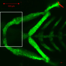
This series of images from late embryonic stages--2 days post-fertilization (dpf)--to late larval stages—21 dpf--shows the appearance, overall anatomical arrangements, and gross morphology of cartilage and bone elements of the ventral jaw skeleton (Arch 1) of the developing zebrafish craniofacial apparatus. In most images, we use the zc81Tg transgenic zebrafish, with fluorescent chondrocytes, to visualize cartilage and Alizarin red to visualize bone. In one image (3 dpf), we use the sp7:EGFP transgenic zebrafish, with fluorescent osteoblasts, to visualize bone formation prior to Alizarin red staining of bone matrix. Images are taken from both ventral (with lateral to top) and lateral views.
|
Info
Click linked text in this column to see full page image(s)
|
Image
Please hold mouse over images to see annotations.
Click on magnifying icon to view large image. |
Description
Click on an underlined term to see its definition and link to related pages
|
|
|
zc81Tg
Alizarin red
|
|
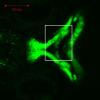
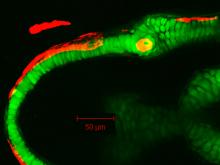
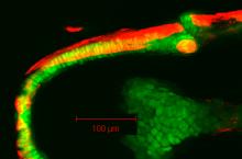
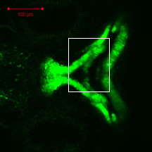
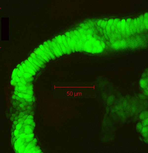
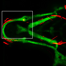
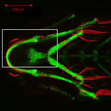
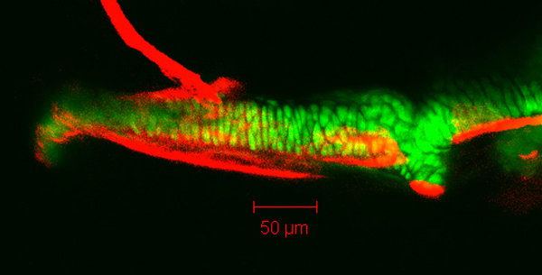
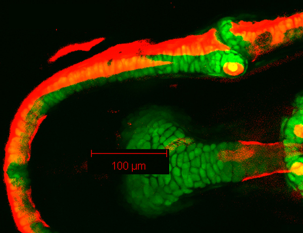
By 55hpf, Meckel’s cartilage (Mk) and the palatoquadrate (pq), derived from the first pharyngeal arch, are becoming distinct elements, with a nascent joint (j) region apparent that is GFP-negative. Alcian blue staining of matrix is observed in these developing cartilages at this timepoint (data not shown). The retroarticular process (rap) of Meckel’s cartilage projects ventroposteriorly and the pterygoid process (ptp) of the palatoquadrate projects dorsoanteriorly. GFP-positive cells are sparse in the medial mandibular symphysis (sym).
|
|
|
zc81Tg
Alizarin red
|
|
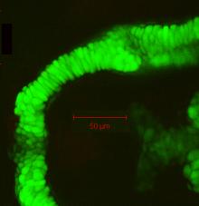
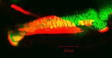
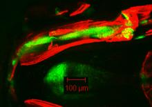
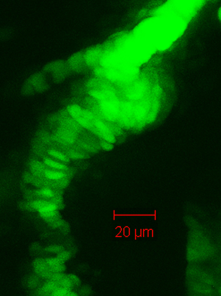
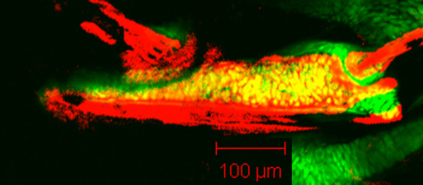
By 78hpf, Meckel’s cartilage (Mk) has grown considerably from that seen at 55 hpf, and a distinct, GFP-negative joint (j) region separates Meckel’s cartilage from the palatoquadrate (pq). Typically, chondrocytes of Meckel’s cartilage appear pseudostratified at this timepoint, not quite spanning the mediolateral width of the element. The retroarticular process (rap) of Meckel’s cartilage projects ventroposteriorly. GFP-positive cells span the medial mandibular symphysis (sym), although those near the midline have relatively lower expression. |
|
|
sp7:EFP
Alizarin red
|
|
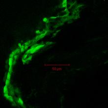
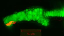
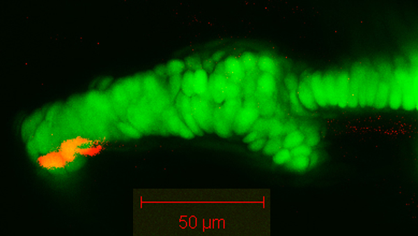
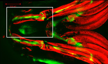
By 78 hpf, the sp7 transgene demonstrates GFP-positive osteoblasts of the dentary (d) that have appeared along the anterolateral surface of Meckel’s cartilage (mouse over to see DIC image of Meckel’s cartilage), although no Alizarin red staining of bone matrix is visible at this timepoint. Expression of the transgene is also visible in ectodermal cells of the jaw epithelium (skin). Of interest to EvoDevo (evolutionary developmental biology) research: |
|
|
zc81Tg
Alizarin red
|
|
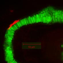
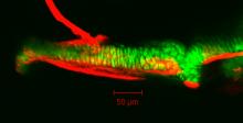
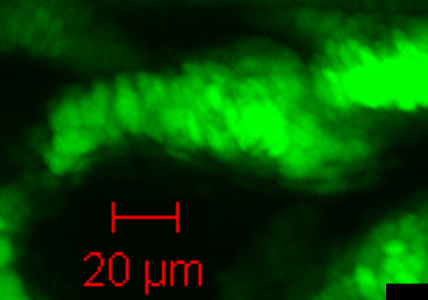
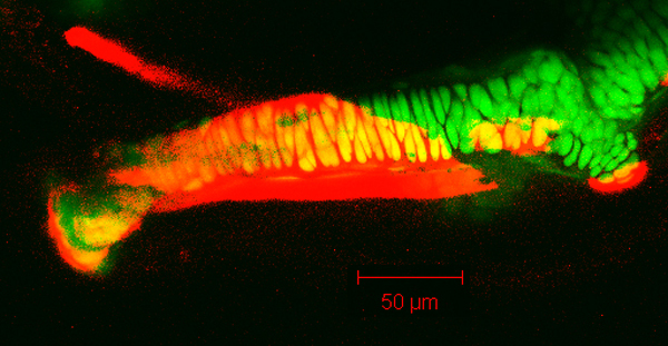
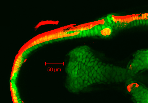
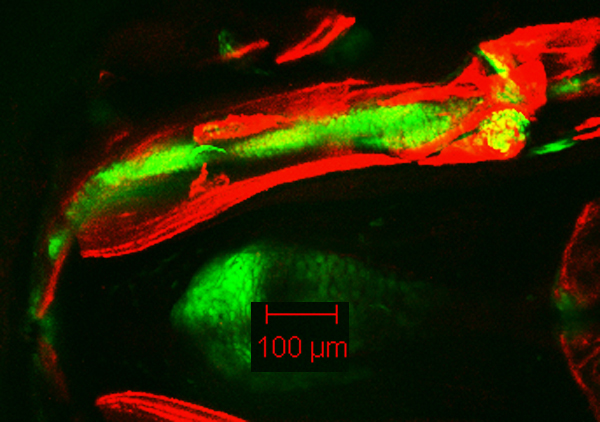
By 86 hpf, Meckel’s cartilage (Mk) has increased in size from that seen at 78 hpf. Most chondrocytes of Meckel’s cartilage now span the mediolateral width of the element, which is consistent with growth by cell intercalation between 78 hpf and 86 hpf. Alizarin red staining also reflects the appearance of the dentary (d) along the ventral aspect of the anterolateral surface of Meckel’s cartilage. The retroarticular process (rap) of Meckel’s cartilage projects ventro-posteriorly from its articulation with the palatoquadrate (pq).
|
|
|
zc81Tg
Alizarin red
|
|
By 98 hpf, Meckel’s cartilage (Mk), the palatoquadrate (pq), the dentary (d), and the maxilla (mx) are visible. The dentary follows the contour of Meckel’s cartilage posterolaterally, projecting away from the cartilage ventrally. GFP-positive chondrocytes span the mandibular symphysis (sym), where cells are positioned ventrally relative to the trajectory of the main body of Meckel’s cartilage.
|
|
|
zc81Tg
Alizarin red
|
|
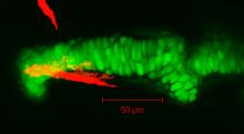
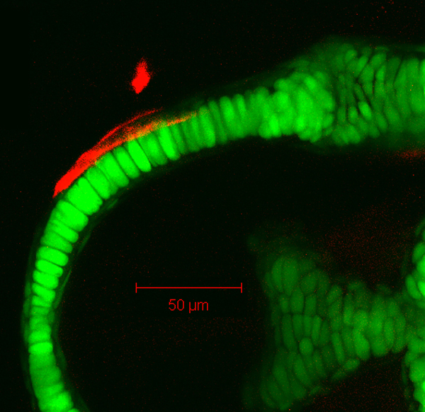
By 105 hpf, the retroarticular (ra) bone can be seen in the most distal perichondrium of the retroarticular process of Meckel’s cartilage.
|
|
|
zc81Tg
Alizarin red
|
|
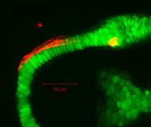
By 149 hpf, Meckel’s cartilage (Mk) has grown in size from that seen at 105 hpf. Note the appearance of chondrocyte doublets along the body of the Meckel’s cartilage in the lateral view, which is consistent with the idea that growth along the length of the cartilage is also accomplished by division of resident chondrocytes. Also visible in these images are the palatoquadrate (pq), dentary (d), maxilla (mx), retroarticular bone (ra), anguloarticular (aa), and quadrate (q). The dentary follows the lateral contour of Meckel’s in both anterior and posterior directions, projecting away from the cartilage along its posteroventral aspect. The anguloarticular appears in the posterolateral perichondrium of Meckel’s cartilage. Cells near the mandibular symphysis (sym) are positioned ventrally relative to the trajectory of the main body of Meckel’s cartilage.
|
|
|
zc81Tg
Alizarin red
|
|
By 199 hpf, Meckel’s cartilage (Mk) can has grown considerably from that seen at 149 hpf. Also visible here are the palatoquadrate (pq), dentary (d), maxilla (mx), retroarticular bone (ra), anguloarticular (aa), and quadrate (q). The dentary has now almost completely covered the lateral contour of Meckel’s cartilage in both anterior and posterior directions, including the ventrally displaced portion of Meckel’s cartilage near the mandibular symphysis (sym). The dorsal extension of the dentary can be seen to articulate with the ventral aspect of the maxilla. The anguloarticular has extended further in the perichondrium of Meckel’s cartilage both posteriorly and anteriorly, its anterior-most aspect now medial to the overlying dentary. Of interest to EvoDevo (evolutionary developmental biology) research: |
|
|
zc81Tg
Alizarin red
|
|
By 247 hpf, Meckel’s cartilage (Mk) and the palatoquadrate (pq), dentary (d), maxilla (mx), retroarticular bone (ra), anguloarticular (aa), and quadrate (q) maintain their relative positions with some growth from that seen at 199 hpf. The lateral contour of Meckel’s cartilage is now completely covered by the dentary and the anguloarticular, with the dentary lateral to the anguloarticular. Bone is beginning to surround Meckel’s cartilage anteriorly near the mandibular symphysis (sym).
|
|
|
zc81Tg
Alizarin red
|
|
By 338 hpf, Meckel’s cartilage (Mk) and the palatoquadrate (pq), dentary (d), premaxilla (pm), maxilla (mx), retroarticular bone (ra), anguloarticular (aa), and quadrate (q) are visible. The dentary has now covered the circumference of Meckel’s cartilage near the mandibular symphysis (sym), which is unlike that seen along the rest of the length of Meckel’s cartilage, where it only extends along one edge (i.e., laterally). Meckel’s cartilage now is displaced dorsally at the mandibular symphysis (data not shown). Note the extensive articulation between the dentary and maxilla, as well as the developiing articulation between the anguloarticular, quadrate, and retroarticular in the jaw joint.
|
|
|
zc81Tg
Alizarin red
|
|
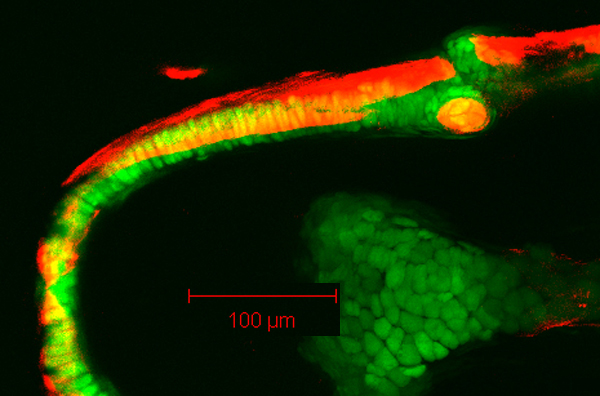
By 514 hpf, Meckel’s cartilage (Mk) has grown considerably from that seen at 338 hpf. Also visible are the palatoquadrate (pq), dentary (d), premaxilla (pm), maxilla (mx), retroarticular bone (ra), anguloarticular (aa), and quadrate (q). The dentary has now grown medially in the dermis along the anterior-posterior length of Meckel’s cartilage, and is found laterally to the anguloarticular posteriorly. Note the complete articulation between bones of the jaw joint, including the quadrate, anguloarticular, and retroarticular.
|

 Movie(s):
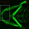


